
Peer Reviewed
What Are These Multiple Papules on a 2-Month-Old’s Face?
Correct answer: D. Neonatal cephalic pustulosis
Neonatal cephalic pustulosis (NCP) is a pediatric skin condition that affects around 10% to 66% of all infants.1 It appears within the first month of life and can persist for up to 6 months.2 Due to the delicate and biochemically fragile nature of neonatal skin, there is a wide variety of dermatologic conditions that can arise early in life.1 Unfortunately, this means that an NCP diagnosis can be overlooked and potentially go untreated. Although it is a benign condition, NCP has the potential to cause significant pruritic discomfort and agitation.
Differential diagnosis. In the case of our 2-month-old infant, a diagnosis of NCP was made on the basis of exclusion by carefully analyzing lesion appearance (ie, no comedones, pustules localized on face/neck, minimal erythema, monomorphic) and his clinical history (onset within first month of life, no maternal complications).2
Miliaria rubra, often referred to as “heat rash,” can manifest within the first 1 to 3 weeks of life due to the obstruction of the immature eccrine glands in the deeper layers of skin.3 Hot environments and physical activity may trigger this condition; thus, it is expected that the lesions will resolve within a few days after removing the stimulus.3 When present, the lesions will appear as small pruritic erythematous papules localized to the neck, groin, and axilla.4 Sometimes, the papules will become pustular, and the condition is named Miliaria pustulosa.2 This condition is self-limited and can be promoted by eliminating inciting factors, such as cooling the skin and avoiding heat exposure.3,5 We excluded this condition in our patient due to infant’s lesions progressive worsening over time, rather than appearing and resolving based on environment. Heat-related lesions also typically appear in areas of occlusion, unlike in our patient, who had many lesions on exposed areas of the face and no lesions in the groin and axilla. Furthermore, the resolution of our infant’s symptoms following ketoconazole treatment indicates an alternative pathology.
Acne neonatorum may arise in the first 4 to 6 weeks of life due to excess hormonal stimulation of sebaceous glands.6 In additon to the infant’s own adrenal glands, this is likely caused by maternal androgens being transferred by the placenta.6 The condition will present as diffuse erythematous pustules and papules. However, unlike NCP, comedones may also be present.7 Rather than using antifungals as in NCP, the treatment of choice is daily cleansing, benzoyl peroxide, topical retinod, or topical antibiotics.7 However, in most cases the acne will self-resolve with time.7 We excluded this conditon due to the lack of comedones. Furthermore, the resolution of our patient’s lesions from an antifungal indicates the pathophyiolsogy was likely not hormonal.
Erythema toxicum neonatorum is a common but benign condition that appears within the first 4 days of life and then quickly resolves within 1 to 2 weeks.8 The exact etiology is unknown, but it likely involves a complex interplay between autoimmunity, hair follicles, and microbes.9 The lesions appear as small papules/vesicles/pustules (1-2 mm) surrounded by a large blotchy erythematous base (1 cm). Although commonly found on the face and trunk, they can be found on any hair-bearing surface.5 Smears of vesicular fluid show a predominance of eosinophils.2 Although benign and self-limited, gentle cleaning of purulence and scale may help resolution.5 We excluded this condition based on the timeline of disease and lack of blotchy erythematous base.
Treatment and management. After NCP was diagnosed, the eruption was treated with 2% topical ketoconazole cream twice a day in accordance with guidelines.10 As expected, the skin cleared after 6 days without scarring (Figures 3 & 4).

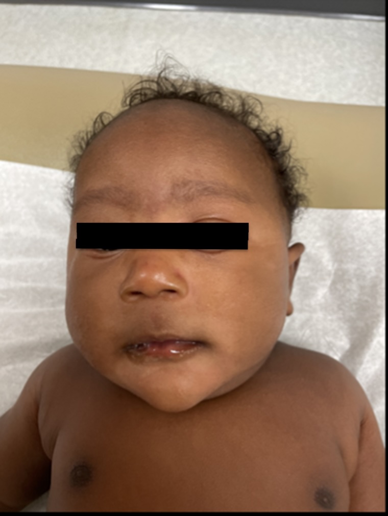
Figure 3. The frontal view of the 3-month-old infant following treatment with 2% topical ketoconazole following diagnosis of NCP, showing significant resolution of pustules and papules from the facial regions of the infant. Erythema has subsided and infant is less irritable. No discoloration is present. Although the mother said improvement was evident in 1 week, this picture was taken 1 month following initiation of treatment.
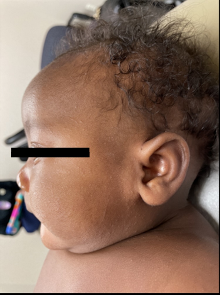
Figure 4. The lateral view of the infant following treatment, showing concomitant resolution of lesions from the scalp, ears, and shoulders. However, a few residual papules remain localized in the temporal region despite treatment.
Outcome and follow-up. No further therapy was continued, and the mother was counseled on skin hygiene for her infant.
Discussion. The exact etiology of NCP lacks consensus. Although some have attributed the condition to hormones, they have also been disputed by studies.1 More recently, the colonization of Malassezia species has been strongly implicated in the pathophysiology of this condition.11 The lesions are known to arise in the first month of life as small and minimally erythematous papules and pustules that are distributed on the head, neck, and chest.5 Notably, unlike acne neonatorum, NCP lacks comedones.5 In clinical practice, the isolation of Malassezia is rarely needed to diagnose NCP.
NCP is often overlooked by physicians despite the significant discomfort it can inflict on newborns. This case study serves to highlight the importance of recording a robust physical examination and history when making a diagnosis. When done sufficiently, it may help to avoid fungal confirmatory testing. Although this condition is relatively benign, treatment with a topical antifungal safely and quickly resolves the skin eruption.
CITATION:
Siddique AS, Gallagher ML. What are these multiple papules on a 2-month-old’s face? Consultant. 2023;63(10):e3. doi:10.25270/con.2023.09.000002.
Received February 21, 2023. Accepted May 9, 2023. Published online September 12, 2023.DISCLOSURES:
The authors report no relevant financial relationships.ACKNOWLEDGEMENTS:
The patient's parents signed patient consent forms allowing Consultant360 to publish the images in this case report.CORRESPONDENCE:
Michelle L. Gallagher, DO, 515 Lake Lansing Road, Suite A-1, Lansing, MI 48912 (docmlg@msu.edu)© 2023 HMP Global. All Rights Reserved.
Any views and opinions expressed are those of the author(s) and/or participants and do not necessarily reflect the views, policy, or position of Consultant360 or HMP Global, their employees, and affiliates.
1. Reginatto FP, Villa DD, Cestari TF. Benign skin disease with pustules in the newborn. An Bras Dermatol. 2016;91(2):124-134. doi:10.1590/abd1806-4841.20164285
2. Ghosh S. Neonatal pustular dermatosis: an overview. Indian J Dermatol. 2015;60(2):211. doi:10.4103/0019-5154.152558
3. Kutlubay Z, Tanakol A, Engýn B, et al. Newborn skin: common skin problems. Maedica (Bucur). 2017;12(1):42-47.
4. Fimiani M, Bilenchi R, Mandato F, et al. Neonatal skin disorders. Neonatology. 2018:2391-2425. doi:10.1007/978-3-319-29489-6_285
5. Chadha A, Jahnke M. Common neonatal rashes. Pediatr Ann. 2019;48(1):e16-e22.
doi:10.3928/19382359-20181206-01
6. Greydanus D, Azmeh R, Demma M, Dickson C, Patel D. Acne in the first three decades of life: an update of a disorder with profound implications for all decades of life. Dis Mon. 2021;67(4):101103. doi:10.1016/j.disamonth.2020.101103
7. Paller AS, Mancini AJ. Hurwitz Clinical Pediatric Dermatology. 6th ed. Elsevier; 2020:11-41.
8. Levy HL, Cothran F. Erythema toxicum neonatorum present at birth. Am J D Child. 1962;103(4):617-619. doi:10.1001/archpedi.1962.02080020630014
9. Marchini G, Nelson A, Edner J, Lonne-Rahm S, Stavréus-Evers A, Hultenby K. Erythema toxicum neonatorum is an innate immune response to commensal microbes penetrated into the skin of the newborn infant. Pediatr Res. 2005;58(3):613-616. doi:10.1203/01.pdr.0000176836.27156.32
10. Jain AK, Morgaonkar M. Acne in childhood: clinical presentation, evaluation and treatment. Indian J Paediatr Dermatol. 2015;16(1):1-4. doi:10.4103/2319-7250.149399
11. Bernier V, Weill FX, Hirigoyen V, et al. Skin colonization by Malassezia species in neonates: a prospective study and relationship with neonatal cephalic pustulosis. Arch Dermatol. 2002;138(2):215-218. doi:10.1001/archderm.138.2.215