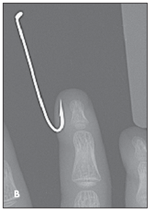
 A 3-year-old boy was at home with his cousin who was preparing for a fishing trip when a fishhook accidentally became lodged in the distal part of the child’s right middle finger (A). There was mild erythema and swelling, with tenderness on palpation. No bleeding or discharge was noted. The patient had full range of motion, with normal sensation and capillary refill. Remaining examination findings were unremarkable. Radiographic views of the affected area confirmed the absence of bony infiltration (B).
The child was afebrile, active, alert, and in no apparent distress. He had no significant medical history; his immunizations were up-to-date.
 There are multiple approaches to dislodging a subcutaneous fishhook.1 Abu Khan, MD, and Mathew Ednick of Brooklyn, NY, emphasize that fishhook removal is unlike other foreign-body removal because advancement through an alternative site is more successful than removal through the initial point of penetration.
The best way to extract a barbed fishhook is the "cut and advance" method:
A wire cutter or trauma shear is used to sever the back end of the hook and shorten the overall length (C).
After the affected area is anesthetized and cleansed thoroughly, the fishhook is slowly advanced forward through a different intact site in the skin. This method resulted in successful removal of the hook from the patient’s finger (D).
The wound is cleaned with an alcohol( swab, and topical antibiotic lotion is applied. The finger is then wrapped in sterile bandages.
When bone involvement is suspected, imaging studies are needed to verify the hook’s position before it is removed. When there is bone involvement, the hook is pulled back before it is advanced through the soft tissue. Consider tetanus toxoid and prophylactic antibiotics in patients whose immunization status is unknown or not up-to-date.


