
How Would You Treat These Progressive Worsening Unhealed Skin Lesions?
A 90-year-old man came to emergency department (ED) with a few days history of redness and swelling at back of the left leg. He denied any insect or spider bites. He denied any fever or chills. The patient had history of compensated congestive heart failure, diet-controlled diabetes, ulcerative colitis in remission without any medicines for 5 years, and history of septic shoulder arthritis 4 years prior.

Figure: Lesion after second incision and drainage was performed.
Physical examination. Physical exam showed a 1 cm area on back of left leg was indurated, red, and tender on palpation. No rashes were noted on the body. The rest of the physical exam was unremarkable with no significant heart murmur. He was treated with oral cephalexin.
SECOND VISIT
Nine days later, patient visited the ED again. This time the induration was bigger (about 3 cm in size) and very tender on palpation. An incision and drainage was performed. Wound culture was showed Klebsiella pneumoniae, sensitive to amoxicillin/clavulanic acid, cefoxitin, cephalothin, ciprofloxacin, moxicloxacin, septra, tetracycline, and doxycycline.
Laboratory tests. Basic metabolic panel was as unremarkable. Complete blood count showed mild stable anemia, normal white cell count. Blood cultures were negative.
SUBSEQUENT VISITS
The patient visited the ED a few times within 1 month for the same reason. A second incision and drainage was performed again (Figure) as another indurated tender skin lesion developed adjacent to initial wound. Antibiotics were changed 4 times and included cephalexin, clindamycin, ciprofloxacin, and sulfamethoxazole/trimethoprim.
The patient had developed at least 7 indurated lesions on left leg and thigh and 2 lesions on right leg. He was evaluated by infectious disease and dermatology specialists.
A CT scan of the left leg revealed diffuse subcutaneous edema along with foci of subcutaneous emphysema.
The patient was admitted to hospital for progressive worsening unhealed skin lesions on left leg with failure to oral antibiotic. He was treated with an intravenous antibiotic.
What's your diagnosis?
Answer on next page
Answer: Pyoderma Gangrenosum
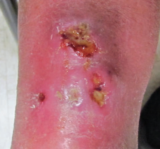
Figure: Lesion after second incision and drainage was performed.

Figure 2: Healed lesion after injectable and topical corticosteroid therapy.
The dermatology consultant suspected that the lesion was pyoderma gangrenosum (PG), and the antibiotics were stopped. The patient was discharged home after 1 dose of betamethasone via intramuscular injection.
The lesion showed some improvement after the betamethasone injection; however, it had worsened 2 weeks later, at which time the patient received a second betamethasone injection, along with topical desoximetasone cream, 0.25%. The lesion on the left leg then completely healed (Figure 2), and all of the other indurated lesions on the legs resolved a few weeks later.
DISCUSSION
PG is a rare neutrophilic skin disorder characterized by painful and necrotic ulceration. Contrary to its name, PG is neither an infectious nor a gangrenous condition.
The typical presentation of PG is an inflammatory papule or pustule that progresses to a painful ulceration. The variants of PG are ulcerative, bullous, pustular, vegetative, and peristomal forms.1 It commonly occurs on the lower extremities and typically affects persons between the ages of 30 and 60 years. More than 50% of patients with PG have associated underlying systemic disease such as inflammatory bowel disease, a hematologic disorder, or various forms of inflammatory arthritis.1
PG can be very challenging to diagnose. The initial presentation of PG is similar to a pyogenic infection, making I&D necessary in some cases. However, the operative approach may exacerbate the lesion due to the pathergy effect.2
In our case, the patient was 90 years old. Even though he had history of ulcerative colitis, his history of shoulder septic arthritis led to more clinical suspicion for an infectious etiology rather than ulcerative colitis. His initial presentation mimicked an abscess, and I&D led to an unhealed wound due to pathergy.
The diagnosis of PG mainly depends on clinical suspicions and the exclusion of other inflammatory or ulcerative cutaneous disorders. No specific laboratory or histopathologic findings confirm the diagnosis. Therefore, a high index of clinical suspicion is very important.
Management of PG depends on the severity and extent of the lesions and underlying disease. Local wound care with moist dressings, and avoidance of aggressive debridement, are generally recommended.3 Topical corticosteroids or topical tacrolimus are used for mild localized PG. More extensive disease or PG that has failed local intervention can be managed with systemic glucocorticoid therapy. Cyclosporine with or without systemic glucocorticoids may be used as an alternative first-line treatment, and immunomodulating agents can be used as second-line or adjunctive therapy.3
References:
- Ahronowitz I, Harp J, Shinkai K. Etiology and management of pyoderma gangrenosum: a comprehensive review. Am J Clin Dematol. 2012;13(3):191-211.
- Wong WW, Machado GR, Hill ME, Pyoderma gangrenosum: the great pretender and a challenging diagnosis. J Cutan Med Surg. 2011;15(6):322-328.
- Callen JP, Jackson JM. Pyoderma gangrenosum:an update. Rheum Dis Clin North Am. 2007;33(4):787-802.