Extravasation of Intravenous Fluid in a Preterm Neonate
An infant was born via cesarean delivery to a 29-year-old, gravida 3, para 2 mother at 31 weeks’ gestation. The pregnancy had been complicated by preterm labor and preeclampsia.
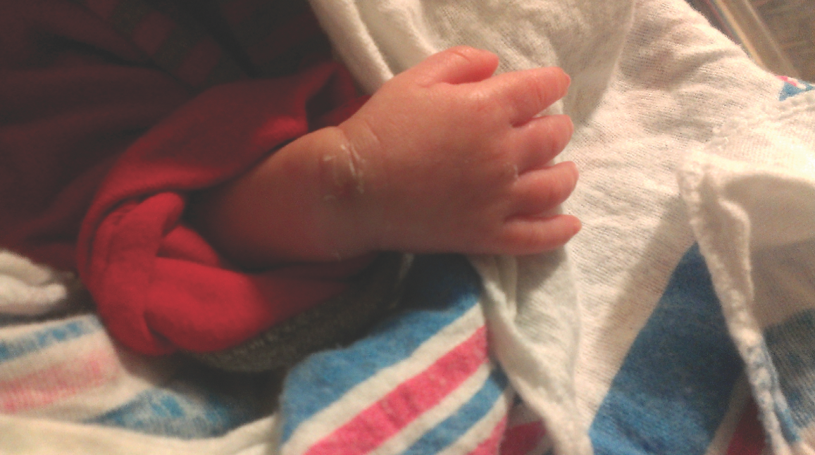
The infant needed mild respiratory support and had an uneventful course in the neonatal intensive care unit. At 29 days of age, however, the neonate developed apnea. Because of suspicion of sepsis, enteral nutrition was stopped, and intravenous total parenteral nutrition was started. After 12 hours of receiving peripheral parenteral nutrition, the infant developed extravasation on the right wrist (Figure 1).

Figure 1. Photograph of the infant’s right wrist showing extravasation of intravenous fluid after having received peripheral parenteral nutrition but before treatment with hyaluronidase.
Subcutaneous hyaluronidase was given circumferentially around the cannula insertion site within 1 hour of extravasation. Bacitracin was applied, and the wound was covered with a sterile dressing.
The wound healed very well without scarring or contracture of the wrist or digits. After 9 days, there was a small, barely noticeable area of hyperpigmentation of the superficial skin at the site of entry of the cannula (Figures 2 and 3).
Discussion
Intensive care of preterm infants requires the insertion of intravascular catheters for parenteral nutrition. Extravasation of fluid, a common complication of peripheral intravenous therapy, can cause significant and long-lasting sequelae in these already compromised patients, and 70% of such injuries occur in infants of 26 weeks’ gestation or less.1 Extravasation injuries are a potentially serious consequence of all intravenous therapy, but they generally are associated with osmotically active solutions containing 10% glucose, calcium chloride, or calcium gluconate, which are standard in neonatal hyperalimentation.1-3

Figures 2 and 3. Photographs of the infant’s wrist 9 days after hyaluronidase treatment. The wound had healed very well without scarring or contracture of the wrist or digits.

No randomized controlled trials to date have demonstrated the effectiveness of the available modes of post-extravasation intervention in reducing injury or scarring, and information about management largely is limited in the literature to case reports. Therefore, no standardized treatment exists for extravasation injuries. Leaving the wound exposed, infiltration with hyaluronidase, and application of occlusive dressings are used with equal frequency, although leaving the wound exposed to the air is not considered optimal treatment for these injuries.1
Infiltration with hyaluronidase is an invasive procedure that is recommended in standard texts,4 and case reports have been published showing its use.5 Hyaluronidase modifies the permeability of connective tissue by hydrolyzing hyaluronic acid, a polysaccharide found in the intercellular ground substance of connective tissue. This temporarily decreases the viscosity of the cellular cement and promotes diffusion of injected fluids or of localized transudates or exudates over a larger surface area, thus facilitating their absorption.
In the absence of hyaluronidase, large subcutaneous infiltrates spread and resolve very slowly. They may exert prolonged pressure on local tissues and surrounding structures, potentially inducing injury, necrosis, and scarring. These are of particular concern when the injury is over a joint because of the possibility that scar contractures could limit joint motility. Further research is needed to help prevent these injuries, and to determine which is the best treatment to aid healing and reduce scarring.
The outcome in this case was excellent despite the location of this large injury over the wrist.
References:
1. Wilkens CE, Emmerson AJ. Extravasation injuries on regional neonatal units. Arch Dis Child Fetal Neonatal Ed. 2004;89(3):F274-F275.
2. Heckler FR, McCraw JB. Calcium-related cutaneous necrosis. Surg Forum. 1976;27(62);553-555.
3. Upton J, Mulliken JB, Murray JE. Major intravenous extravasation injuries. Am J Surg. 1979; 137(4):497-506.
4. Harding S. Procedures and iatrogenic disorders. In: Rennie JM, ed. Rennie & Robertson’s Textbook of Neonatology. 3rd ed. Philadelphia, PA: Churchill Livingstone Elsevier; 1999:1261-1289.
5. Davies J, Gault D, Buchdahl R. Preventing the scars of neonatal intensive care. Arch Dis Child Fetal Neonatal Ed. 1994;70(1):F50-F51.
Dr Khan, Dr Rizvi, and Dr Malik are in the Department of Pediatrics at the Rutgers Robert Wood Johnson Medical School (RWJMS) in New Brunswick, New Jersey. Dr Wienberger is a professor of pediatrics and chief of the Neonatology Division, Dr Puvabanditsin is an assistant professor of pediatrics, and Dr Hegyi is program director of the Neonatology Division in the Department of Pediatrics at RWJMS.


