Images of Malignancy
 Chondrosarcoma
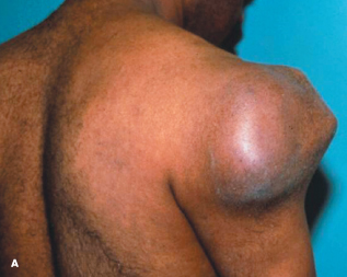
A firm mass projected from the deltoid muscle region of a 24-year-old man’s right shoulder. The patient stated that the mass, which had been present “for years,” had always been firm and immobile. Three months before seeking evaluation, the patient’s upper right arm and shoulder became painful and the mass grew more prominent (A).
A radiograph demonstrated a 10-cm lobulated and calcified mass centered in the metaphyseal region of the proximal humerus. The lobulations contained the arc and ring pattern typical of a cartilaginous neoplasm (B). A nuclear medicine bone scan and a full-body skeletal survey demonstrated no additional lesions. The mass was resected; the proximal and mid humerus were removed as well (C).
Examination of a specimen from a frozen section of the neoplasm revealed a low-grade chondrosarcoma, which arose within an existing osteochondroma. The average size of a chondrosarcoma that arises from an enchondroma, or a benign growth of cartilage within bone, is 5 to 6 cm or greater. Clinically, malignant transformation is suspected when:
•The lesion enlarges rapidly.
•The patient complains of pain.
•An ultrasound or MRI scan shows the lesion’s cartilage cap is greater than 1 to 2 cm.
The treatment of chondrosarcoma is determined by the stage of the neoplasm; the prognosis largely depends on the tumor’s grade. Resection is the treatment of choice for localized tumors; chemotherapy and radiation therapy have no role if, as in this case, the tumor is resectable.
An osteoarticular allograft was used to reconstruct the patient’s
humerus. He will undergo follow-up MRI examinations at 3 and 6 months, and 1 and 2 years after tumor resection. Evaluations will then be scheduled every 2 to 4 years and may continue for 10 years after resection.

(Case and photographs courtesy of Drs John Whyte and Douglas Bell.)

Renal Cell Carcinoma Metastases to the Lung
A 2-month history of cough sent a 62-year-old woman for medical evaluation. The patient denied fever, chills, and rigors but reported seeing streaks of blood in her sputum during the past month. The patient had undergone a right nephrectomy 4 years earlier for renal cell carcinoma.
A chest film disclosed multiple nodules in both lungs. A biopsy of the nodules confirmed metastatic renal cell carcinoma. There was no evidence of endobronchial lesions on bronchoscopy.
The roentgenographic pattern of multiple metastatic lung nodules varies from diffuse micronodular shadows to large, well-defined masses, or “cannonballs.” When first seen, approximately 30% of patients with renal cell carcinoma have distant metastases. However, as in this case, a considerable amount of time may elapse between the initial discovery of the renal tumor and the identification of metastases. Lung metastases with renal cell carcinoma have a poor prognosis, and only palliative treatment can be tried.
The cannonball lesions caused the patient’s persistent cough and hemoptysis. Extensive bone metastases were discovered as well; the patient was referred for
radiation therapy.

(Case and image courtesy of Drs Mahesh Duggal, Krishna Badhey, and Arunabh.)

Cutaneous T-Cell Lymphoma
Three months ago, a 50-year-old man who was otherwise in good health noticed a hard, round nodule on his left arm. Within 2 months, similar nodules appeared all over his trunk, head, arms, and legs. The reddish purple lesions, less than 2 cm in diameter, were painless and slightly pruritic.
The patient was hospitalized, at which time neither lymphadenopathy nor organomegaly was noted. Results of a complete blood cell count, biochemical studies, and urinalysis were normal, and an ECG, a chest film, and CT studies of the thorax and abdomen revealed no abnormalities. Serologic tests were negative for hepatitis B and C viruses, Epstein-Barr virus, cytomegalovirus, HIV, human T-cell leukemia/lymphoma-1 and -2 viruses, rickettsiae, and Treponema pallidum. Biopsy results of bone marrow aspirate were inconclusive.
The diagnosis was obtained from histopathologic examination of a skin biopsy specimen: high-grade cutaneous T-cell lymphoma. The patient was placed on a chemotherapy regimen consisting of cyclophosphamide, doxorubicin, vincristine, and prednisone (CHOP), and a remarkable regression of the skin lesions was evident after 40 days.
(Case and photograph courtesy of Drs Nicholas Pritsivelis, Haralampos Milionis, and Moses Elisaf.)

Glioblastoma Multiforme
A 57-year-old man was brought to the emergency department with severe bifrontal headache, which he had had for 3 weeks. Family members reported that the patient exhibited episodes of confusion and loss of recent memory since the onset
of the headache.
The patient denied nausea and vomiting, fever, visual disturbances, seizures, and weakness of the extremities. The physical examination revealed bilateral papilledema; all other findings, including those regarding higher mental function, were normal. Routine laboratory test results and chest film findings were within normal limits.
A CT scan with contrast demonstrated a large heterogeneous mass with peripheral enhancement in the left frontal lobe, with surrounding edema and midline shift (A). Intravenous corticosteroids were given, and the patient responded with mild clinical improvement. An MRI showed the same mass, edema, and midline shift. A subtotal resection of the tumor was performed; no focal deficits resulted.
The pathologic examination of the lesion confirmed the clinical suspicion of glioblastoma multiforme. Characteristic areas of hypercellularity and cellular atypia (B, double arrows) and central necrosis (B, single arrows) were seen. Vascular and endothelial cell proliferation (C, arrow) was also detected in a glioblastomatous background.
The patient is being treated currently with cranial irradiation.

(Case and images courtesy of Drs Thomachan Kalapura and Viswanatha Kurukundha.)


